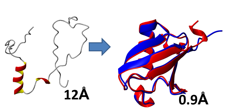
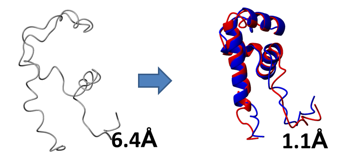
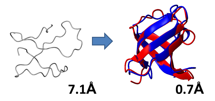
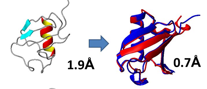
Protein model accuracy before and after CS-GAMDy refinement.
Starting models are shown on the left. β-strands are colored blue, [Symbol]-helices are colored red and yellow, coil regions are colored gray. Alignments of refined models (red) with the reference models (blue) are shown on the right. Numbers represent the RMSD difference between the models and the reference structure (CA RMSD of non-coil regions) before and after refinement.
(A) Distorted model of ubiquitin (reference PDB ID: 1UBQ):
(B) Distorted model of Q5E7H1 (reference PDB ID: 2JVW):
(C) Distorted model of CSPA (reference PDB ID: 1MJC):
(D) Comparative model of cg2496 (reference PDB ID: 2KPT, template PDB ID: 2KW7, sequence ID: 24%):

(E) Comparative model of ubiquitin (reference PDB ID: 1UBQ, template PDB ID: 1IYF, sequence ID: 30%):
(F) Comparative model of NFU1 homolog (reference PDB ID: 2M5O, template PDB ID: 1TH5, sequence ID: 20%):

|
| |
Problems? Suggestions? Please
contact Wishart group

|